
By David Quig, PhD
In 2018 the CDC reported that in the US 60% of adults and 20% of children and adolescents were obese or overweight (BMI>25).1 Cardiometabolic disorders have increased at disturbing rates, and visceral fat deposition is a central factor. Lifestyle choices regarding diet and physical activity are certainly in play. However, modifications of caloric intake and energy balance alone appear insufficient to overcome the extensive influence of the gastrointestinal (GI) microbiota on the initiation and progression of metabolic and cardiovascular complications. The GI microbiota are pivotal factors in the regulation of chronic low-grade inflammation, glycemic control, endothelial dysfunction, and associated cardiometabolic comorbidities. Research-based advances in diagnostic testing have led to the availability of highly focused laboratory tests to evaluate GI dysbiosis and serum-based cardiometabolic risk factors.
Obesity has increased at an alarming rate among adults, and excess adiposity is also more prevalent among children and adolescents.2 Obesity is associated with cardiometabolic risk factors, including dysglycemia, dyslipoproteinemia, sterile endotoxemia, and low-grade inflammation. Specifically, abdominal visceral fat (VAT) is a strong independent predictor of mortality, and it has a central role in pathogenesis of metabolic syndrome (MetS) and coronary artery disease (CAD).3 The extent of fat deposition in the three primary types of depots is correlated, but VAT is very different from subcutaneous fat with respect to release of fatty acids immediately to the liver and pancreas and pathogenic sequelae.4 Further, macrophages amass more extensively in VAT and release pro-inflammatory cytokines and reactive oxygen species that result in systemic low-grade inflammation, disrupted adipokine metabolism, and insulin resistance.5,6 Low-level systemic elevations of gut microbiota-derived bacterial lipopolysaccharides (LPS) augment infiltration and activation of macrophages and accentuate the release of pro-inflammatory cytokines.5,7,8 Intestinal barrier disruption and high abundance of LPS-rich Proteobacteria (phylum), Enterobacteriaceae (family), and Shigella and Escherichia (genera) are common with inflammatory conditions such as obesity, type-2 diabetes mellitus, inflammatory bowel disease, and high-fat diets.9-11 In contrast, normally abundant commensal species such as Faecalibacterium prausnitzii, Bifidobacterium spp., Eubacterium rectale, and Akkermansia muciniphila are diminished. That loss of commensals begets increased inflammation and intestinal permeability (paracellular).12,13 Such an inflammatory microbiota profile is readily detected and characterized via a highly focused PCR-based dysbiosis test.11
An abundant and diverse microbial community is essential for the maintenance of the intestinal barrier function and normoglycemia. Adequate intake of soluble fiber and GI eubiosis provides a balanced abundance of short-chain fatty acids (SCFA) that regulate several aspects of host metabolism related to adiposity and MetS.14,15 Butyrate and propionate stimulate proliferation of enteroendocrine L-cells and their release of glucagon-like peptide-1 (GLP-1). GLP-1 regulates post-prandial glycemic control and satiety and attenuates LPS-induced inflammatory pathways in VAT.14 Butyrate and acetate stimulate β-oxidation of fatty acids in lean tissues and the constant secretion by Goblet cells of mucins that constitute the critical mucus barrier gradient.16 The status of colonic SCFAs can be indirectly assessed via a stool specimen.
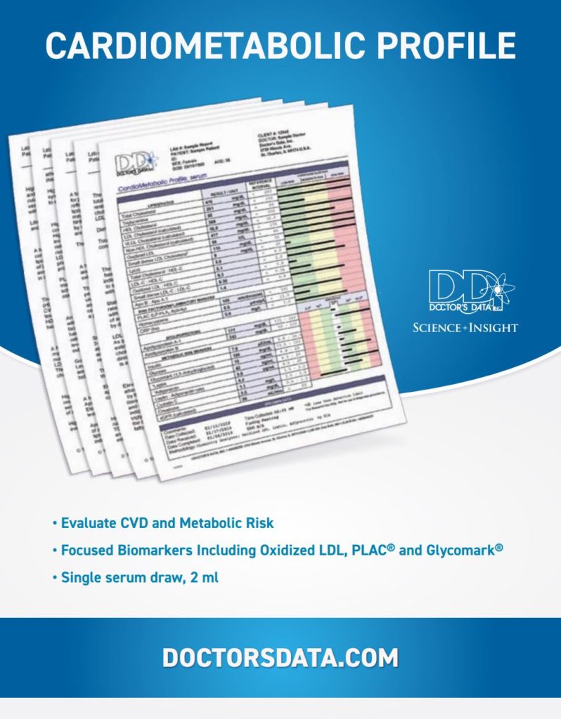
Obesity is associated with an entanglement of cardiometabolic risk factors, including dyslipoproteinemia, dysglycemia, low-grade inflammation, and endothelial and renal dysfunctions. Beyond a dated lipid profile, lipoprotein-related cardiometabolic risk considerations include the serum levels of triglycerides and non-HDL cholesterol, and the levels and ratios of apolipoproteins A-I and B. Also, very important are the levels of the specific low-density lipoprotein (LDL) culprits, oxidized LDL, small dense LDL, and Lp(a), which are independent of total LDL cholesterol. The potential cardiometabolic effects of high-density (HDL) and low-density (LDL) lipoproteins are not well predicted based upon the traditionally measured lipoprotein cholesterol levels.17 Higher apolipoprotein B (apoB) levels, lower HDL-associated apolipoprotein A1 (apoA1), and a higher apoB/apoA1 ratio have strong predictive value with respect to cardiometabolic risk and outcome.18,19 A high level of oxidized LDL (Ox-LDL) is a strong independent risk factor for CAD.20 In the CARDIA study Ox-LDL was also associated with incidence of MetS, and its components of abdominal obesity, hyperglycemia, and hypertriglyceridemia.21 Fasting triglyceride levels are not recognized as an independent risk factor for CAD, but mechanistically very low density lipoprotein (VLDL) triglycerides have a pivotal role in an atherogenic lipoprotein profile. An expanded VLDL triglyceride donor pool initiates formation of atherogenic small dense LDL (sdLDL) particles via the activity of the plasma lipid transfer protein.22 Subsequent hydrolysis of the acquired LDL core triglycerides results in sdLDL. The sdLDL more readily infiltrate the endothelium where the antioxidant-poor sdLDL are susceptible to oxidation by macrophages in the arterial wall.23,24 The LDL-like Lp(a) is also associated with atherogenesis as it is taken up in an unregulated manner by macrophages, and appears to have distinct prothrombotic and proinflammatory properties.25,26

The activity of macrophage-derived lipoprotein-associated phospholipase A2 (PLAC) provides important information regarding vascular inflammation and plaque instability.27 Oxidized lysophospholipids and fatty acids released by PLAC activity on the surface of LDL particles contribute to oxidation of LDL (Ox-LDL) and endothelial inflammation. Concomitantly elevated serum levels of PLAC activity and CRP are associated with greater risk for coronary and stroke events.28
Excess VAT, MetS, and CAD are conditions associated with altered adipokine metabolism. Adiponectin is an abundant adipocyte-derived adipokine that effectuates anti-inflammatory, anti-oxidative, antidiabetic, and vascular protective mechanisms. Excess VAT and MetS are associated with decreased expression of adiponectin, hypoadiponectinemia, and higher levels of opposing leptin.6,29,30 A high leptin-to-adiponectin ratio coupled with elevated hsCRP has strong predictive value for CAD.31,32

Serum insulin and dysglycemia may be assessed directly in the fasted state, and time averaged glycemic control over the past two to three months is indicated by serum levels of HbA1C. However HbA1C levels do not provide information regarding maximal hyperglycemic episodes that are independently associated with CAD and renal damage in type 2 diabetics.33 Beyond HbA1C, an abnormally low serum level of 1, 5-anhydroglucitol (GlycomarkTM) provides specific information regarding daily hyperglycemic episodes over the past one to two weeks. Renal dysfunction associated with cardiometabolic disorders may be thoroughly assessed via serum cystatin C, creatinine and estimated GFR. The level of cystatin C is an excellent inversely related indicator of diminished glomerular filtration, and predictor of subclinical CAD in diabetics.34,35
The prevalence of excessive adiposity among youth and adults will likely have significant long-term cardiometabolic consequences. Early detection of metabolic disruptions and clinical intervention are paramount towards stemming the tide of the epidemic. The GI microbiota and its net metabolism have a central role in the initiation and progression of metabolic and cardiovascular complications. Comprehensive stool analysis inclusive of a highly focused PCR-based dysbiosis test facilitates confident identification of a dysfunctional microbial community. Modern research-based laboratory testing provides clinicians with more specific serum-based metabolic biomarkers that have high predictive value for cardiometabolic disorders. Such a complete kit may also fulfill the monitoring and motivational needs of clinicians.
Click here for complete list of References and related articles, on the next page.


