By Alan R. Gaby, MD
#432
Probiotic Prevents Otitis Media in Children
Sixty-one children (mean age, 3.3 years) living in Spain who were experiencing recurrent acute otitis media received the probiotic organism, Lactobacillus salivarius PS7, orally at a dose of 109 colony-forming units per day for six months. This strain was selected because it had previously been shown to have antimicrobial activity against organisms that can cause otitis media. Compared with the six months before the intervention, there was an 84% decrease in the number of episodes of acute otitis media during probiotic treatment.
Comment: Acute otitis media (inflammation of the middle ear) is usually caused by a bacterial or viral infection, although it may also result from an allergic reaction to foods. Probiotics have the potential to prevent various types of infections by competing with pathogenic organisms and by enhancing immune function. The probiotic used in this study was selected specifically because of its capacity to inhibit the growth of otopathogens, so one cannot assume that other probiotic strains would have a similar effect. In addition, because there was no placebo group in this study, it is possible that some or all of the observed improvement was due to a placebo effect or to age-related spontaneous remission. The product used in this study does not appear to be commercially available at this time.
Other interventions that may help prevent otitis media include restricting sugar intake, identifying and avoiding allergenic foods, and regular oral use of xylitol chewing gum or syrup.1
Cardenas N, et al. Prevention of recurrent acute otitis media in children through the use of Lactobacillus salivarius PS7, a target-specific probiotic strain. Nutrients. 2019;11:E376.
Probiotics Prevent Clostridium difficile Infection in Patients Receiving Antibiotics
A recent Cochrane review pooled the results of 39 randomized controlled trials that examined whether supplementing with probiotics can prevent the development of Clostridium difficile infection in patients receiving antibiotics. In the pooled analysis, the incidence of C. difficile infection was 60% lower in patients receiving probiotics than in those receiving placebo or no treatment (1.5% vs. 4.0%; p < 0.001).

Comment: C. difficile is a Gram-positive bacterium that is a common cause of antibiotic-associated diarrhea and pseudomembranous colitis. C. difficile infection occurs most often in frail elderly hospitalized patients but has also been seen in previously healthy individuals. The infection usually manifests as mild-to-moderate diarrhea, but severe colitis culminating in colectomy or death may also occur. In recent years, there has been an increase in the incidence of C. difficile infection. Increases in disease severity and mortality rates have also been observed, apparently because of the emergence of a more virulent strain of the organism.2 C. difficile infection is usually treated with vancomycin or metronidazole. The infection recurs in approximately 20% of patients after treatment with these antibiotics. Probiotics may prevent the development or reduce the recurrence rate of C. difficile infection by competing with C. difficile for nutrients and colonization sites in the intestinal tract, by producing compounds that inhibit the growth or decrease the virulence of C. difficile, or by producing compounds that neutralize C. difficile-associated toxins.
Although probiotic preparations are generally well tolerated, in rare cases sepsis due to Saccharomyces fungemia or Lactobacillus bacteremia has occurred after oral administration of the respective probiotic agent. Risk factors for the development of this complication include being debilitated or immunosuppressed, having a damaged gastrointestinal barrier, receiving broad-spectrum antibiotics, and having a central venous catheter.
Goldenberg JZ, et al. Probiotics to prevent Clostridium difficile infection in patients receiving antibiotics. JAMA. 2018;320:499-500.
Thiamine Reduces Mortality in Patients with Septic Shock

A retrospective cohort study was conducted on 123 patients with septic shock who received intravenous thiamine within 24 hours of hospital admission (median, 6.4 hours; range, 3.8-11 hours) and 246 matched controls who did not receive thiamine. Two-thirds of the patients received 500 mg of thiamine every eight hours for three days; the others received 100-400 mg (apparently every 8 hours for 3 days). Compared with no thiamine, thiamine treatment was associated with more rapid lactate clearance (which is a predictor of increased survival) and a significant 33.4% reduction in 28-day mortality.
Comment: Septic shock is a serious condition that has a mortality rate of 40-50%. Critically ill patients are frequently deficient in thiamine, and in these patients the presence of thiamine deficiency is associated with an increased risk of death. In the October 2017 issue of the Townsend Letter, I discussed the work of Dr. Paul Marik and coworkers, who administered vitamin C, hydrocortisone, and thiamine intravenously to 47 patients with septic shock. As compared with similar patients who did not receive this treatment, those given vitamin C, hydrocortisone, and thiamine had a 79% reduction in the mortality rate. Previous research had provided evidence that intravenous vitamin C by itself can reduce the death rate in patients with severe sepsis. The results of the present study suggest that thiamine was an important component of Marik’s protocol, and that it may have enhanced the benefits of vitamin C.
Woolum JA, et al. Effect of thiamine administration on lactate clearance and mortality in patients with septic shock. Crit Care Med. 2018;46:1747-1752.
Does Cow’s Milk Cause Iron-Deficiency Anemia?
Of 51 children under four years of age (median, 1.4 years) in Taiwan who had iron-deficiency anemia, seven (13.7%) had cow’s milk protein allergy. Four of those seven children had occult blood in their stool. All seven patients recovered from iron-deficiency anemia within seven months of avoiding cow’s milk and receiving iron supplementation. All four children with positive occult blood in their stool became negative after 1.5 months of cow’s milk avoidance.
Comment: Allergy to cow’s milk was implicated as early as the 1960’s as a common cause of both occult and gross gastrointestinal bleeding in infants; and it appears to be an important contributing factor to anemia in that age group. The adverse effect of fresh pasteurized cow’s milk appears to be much more pronounced than that of commercial milk-based formulas. The results of the present study should remind us to consider cow’s milk sensitivity in children with unexplained iron-deficiency anemia.
Lai FP, Yang YJ. The prevalence and characteristics of cow’s milk protein allergy in infants and young children with iron deficiency anemia. Pediatr Neonatol. 2018;59:48-52.
Is Cow’s Milk “Mucus Forming’?
Twenty-six men and 82 women who experienced symptoms of excessive nasopharyngeal mucus secretion, who had no history of intolerance to cow’s milk or soy protein, and who had negative skin prick tests to milk and soy, consumed a dairy-free diet for six days. During the last four days on this diet, they were randomly assigned to consume, in double-blind fashion, a daily cow’s milk-based or soy-based milkshake (350 ml per day). During the first three days (no dairy in either group), both groups experienced a significant reduction in the mean severity of mucus secretion. On the last day of the study, the amount of nasopharyngeal mucus secretion was significantly less in the dairy-free group than in the group that consumed dairy. The effect size was 0.55 (medium).
Comment: There is a widespread belief that drinking cow’s milk increases mucus formation, but there has been little scientific support for that idea. In the present study, consumption of a dairy-free diet significantly decreased mucus secretion in adults with symptoms of excessive nasopharyngeal mucus secretion. Cow’s milk is one of the most common food allergens, and mucus secretion is one of the symptoms of an allergic reaction. Therefore, it remains unclear whether there is something inherently mucus-forming about cow’s milk, or whether an increase in mucus production is simply a manifestation of milk allergy.
Frosh A, et al. Effect of a dairy diet on nasopharyngeal mucus secretion. Laryngoscope. 2019;129:13-17.
Does Diet Influence the Risk of Developing Macular Degeneration?

The association between diet and risk of developing aged-related macular degeneration (AMD) was examined in a prospective cohort study of 4,202 participants in the Rotterdam Study who were at least 55 years of age at baseline (mean, 66.6 years) and were free of AMD. During a mean follow-up period of 9.1 years, 754 people developed AMD. Consumption of fish at least twice a week, as compared with less fish consumption, was associated with a significant 24% decrease in the incidence of AMD. Intake of at least 200 g per day of vegetables or intake of fruit at least twice a day was not associated with AMD risk. However, among participants who achieved all three of these dietary patterns (fish, vegetables, and fruit), the incidence of AMD was 42% lower than among those who did not achieve all three dietary patterns.
Comment: In this study, consumption of fish twice a week was associated with a decreased risk of developing AMD. Consumption of abundant amounts of fruits and vegetables were not by themselves associated with a lower incidence of AMD. However, consumption of fruits and vegetables increased the protective effect of fish consumption. While observational studies do not prove causation, the results of this study raise the possibility that eating fish, fruits, and vegetables can help prevent AMD, which is the most common cause of visual loss in elderly people.
de Koning-Backus AP, et al. Intake of vegetables, fruit, and fish is beneficial for age-related macular degeneration. Am J Ophthalmol. 2019;198:70-79.
Whole Grains and Nonalcoholic Fatty Liver Disease
Fifty overweight men (aged 45-70 years) and postmenopausal women were randomly assigned to consume, in double-blind fashion, refined wheat or whole wheat products for 12 weeks. The mean concentration of intrahepatic triglycerides (measured by proton magnetic resonance spectroscopy) increased by 49% in the refined wheat group and increased by 11% in the whole wheat group (p = 0.03 for the difference in the change between groups).
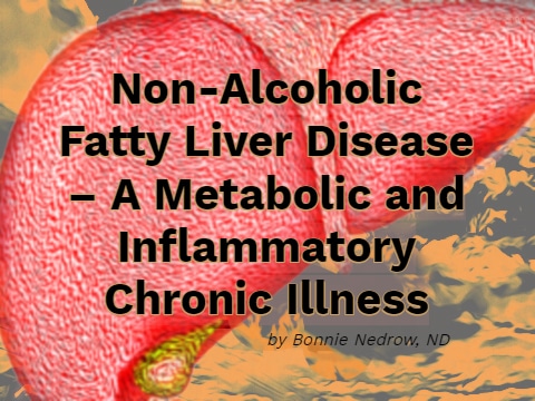
Comment: Hepatic steatosis (excessive accumulation of fat in the liver) is one of the two main features of nonalcoholic fatty liver disease, the other being chronic hepatic inflammation. Hepatic steatosis affects as many as 25% of Americans and 58-74% of obese people. Hepatic steatosis appears to be an independent risk factor for cardiovascular disease and is also associated with increased all-cause mortality. In addition, the presence of hepatic steatosis may increase the progression rate of other liver diseases, such as hepatitis C.
There is evidence that excessive consumption of fructose and sucrose is a major risk factor for the development of hepatic steatosis. The results of the present study suggest that consumption of refined grains is another contributing factor, and that switching to whole grains may decrease the accumulation of fat in the liver. As compared with refined grains, whole grains contain substantially higher amounts of magnesium, copper, choline, betaine, and vitamin E. Each of these nutrients has been shown in animal studies, human trials, or both to be useful for preventing and/or treating fatty liver.
Schutte S, et al. A 12-wk whole-grain wheat intervention protects against hepatic fat: the Graandioos study, a randomized trial in overweight subjects. Am J Clin Nutr. 2018;108:1264-1274.
Selenium and Diabetes
Four hundred ninety-one volunteers (aged 60-74 years) in Denmark (a population with moderately low selenium status) were randomly assigned to receive, in double-blind fashion, 100, 200 or 300 µg per day of selenium (as selenium-enriched yeast) or placebo for two years. Compared with placebo, there were no significant changes in the mean hemoglobin A1c (HbA1c) concentration in any of the three selenium groups, either at six months or at two years. There was a nonsignificant trend suggesting a beneficial effect of selenium on HbA1c levels.

Comment: Concern has been raised that selenium supplementation could increase the risk of developing diabetes. That concern was based on a retrospective analysis of a double-blind study, which found that selenium supplementation (200 µg per day) was associated with a 50% increase in the incidence of new cases of diabetes, compared with placebo, during a follow-up period of 7.7 years (p = 0.05).3 However, when a retrospective analysis reveals an effect of only borderline statistical significance, there is a reasonable likelihood that the finding was due to chance. A meta-analysis of four randomized controlled trials (including the retrospective analysis mentioned above) found that supplementation with 200 µg per day of selenium for 5-12 years nonsignificantly increased the incidence of type 2 diabetes by 9% (p = 0.085).4 Diabetes is not one of the reported manifestations of selenium toxicity in humans. Moreover, in an animal model of type 2 diabetes, selenium supplementation exerted an antidiabetic effect.5 Thus, the small increase in diabetes incidence observed in the meta-analysis may have been due to chance.
The findings from the new study provide evidence that selenium supplementation does not have an adverse effect on blood glucose regulation. However, the new study lasted only two years, so it remains possible that longer-term use of selenium could result in a small increase in diabetes risk.
Stranges S, et al. Effect of selenium supplementation on changes in HbA1c: Results from a multiple-dose, randomized controlled trial. Diabetes Obes Metab. 2019;21:541-549.
REFERENCES
1. Gaby AR. Otitis media. In Gaby AR. Nutritional Medicine, 2nd Edition. Concord, NH, 2017. www.doctorgaby.com, chapter 269.
2. Kelly CP, LaMont JT. Clostridium difficile – more difficult than ever. N Engl J Med. 2008;359:1932-1940.
3. Stranges S, et al. Effects of long-term selenium supplementation on the incidence of type 2 diabetes: a randomized trial. Ann Intern Med. 2007;147:217-223.
4. Mao S, et al. Selenium supplementation and the risk of type 2 diabetes mellitus: a meta-analysis of randomized controlled trials. Endocrine. 2014;47:758-763.
5. Mueller AS, Pallauf J. Compendium of the antidiabetic effects of supranutritional selenate doses. In vivo and in vitro investigations with type II diabetic db/db mice. J Nutr Biochem. 2006;17:548-560.


