by Aristo Vojdani, PhD, MSc, CLS
Abstract: Alzheimer’s disease is a chronic neurodegenerative disease that is the most common cause of dementia. Many people mistakenly believe that genetics is the sole cause of Alzheimer’s, but research accumulated over the years has shown that environmental factors actually have a greater role in the development of the disorder. In this review we show that infectious agents, the toxic chemicals in our environment, even the food we eat daily, and the composition of the bacteria in our bodies can increase the risk of developing Alzheimer’s disease. By their reaction with brain tissue antigens such as amyloid or tau protein, these environmental triggers or antibodies produced against them contribute to the pathophysiology of this disorder. Although statistics show that about 6,000,000 people in the US do suffer from mild cognitive impairment or the advanced stages of Alzheimer’s disease, roughly 47,000,000 people may actually already be in the preclinical stage of the disease. It is important to use reliable and accurate testing such as Alzheimer’s LINX™ to identify the specific triggers that are involved in the development of this disorder, so that medical practitioners can remove these triggers and implement lifestyle modifications to prevent Alzheimer’s disease in the more than 50,000,000 individuals in the preclinical and early stages of the disorder.
For years, like many, I believed that genetics was the cause of Alzheimer’s disease (AD) and other neurodegenerative disorders. Many times I had heard that since my mother got AD at the age of 71 that I was sure to get it as well. However, the latest research indicates that environmental factors such as certain pathogens, toxic chemicals, undigested food antigens, and alterations in gut microbiota can increase the risk of developing AD.1-4 About 6,000,000 people in the US and more than 47,000,000 people worldwide live with AD, and by 2050 this number may exceed 120,000,000 if no effective prevention strategies are used.
AD is divided into two main categories: early and late onset. The early onset of AD occurs between the ages of 30-60 and is almost 100% due to genetics. It is due to mutation in several genes such as APP, PSEN1 and PSEN2 that are involved in the production of amyloid-beta. These mutations are responsible for the accumulation of amyloid plaques and neurofibrillary tangles, neuronal and synaptic loss in the brain. Only about 300,000 out of around 6,000,000 AD sufferers in the US or about five percent suffer from early onset AD. The other approximately 5,700,000 sufferers in the US are afflicted with late onset AD, which occurs after the age of 60.5 Furthermore, according to Brookmeyer et al., in 2017, 46,700,000 Americans, who were in one of the preclinical stages of AD, may or may not develop full-blown AD, depending on their lifestyle.6
Based on this, the changes from normal to full-blown AD are classified into three major stages:
· First stage of AD – Preclinical (46,700,000 in the US)
· Second stage of AD – Mild cognitive impairment (2,500,000 in the US)
· Third stage of AD – AD with dementia (3,500,000 in the US)
ApoE Is Not a Gene for Alzheimer’s Disease
Apolipoprotein E (ApoE) is a lipid carrier, a protein involved in the metabolism of fats in the body. ApoE has been implicated in innate immunity, inflammation, atherosclerosis, and AD.7 There are three common ApoE alleles in the human population: E2, E3 and E4, with ApoE4 present in about 10% of the population. In late-onset AD, the apolipoprotein E4/4 (ApoE4) variant plays a significant role as the risk factor for the disease. Although not every individual positive with ApoE4/4 will get AD, overall the E4 allele is significantly enriched in patients with AD, and E4-carrying patients show heavier amyloid-β plaque load in the brain and earlier disease onset. This is why ApoE was initially identified as an amyloid-β binding protein. Interestingly, substantial evidence has demonstrated a relationship between ApoE and amyloid-β deposition in a dosage-dependent and isoform-specific fashion, with ApoE4 the strongest and ApoE2 the least.7 This function of ApoE is likely linked to receptors, such as the low-density lipoprotein receptor, heparin sulfate, that are expressed on the membranes of microglia cells, to which they tend to bind. In addition to ApoE binding to microglia cells, in the initiation stage of plaque formation, ApoE may participate as an opsonin to enhance the microglia-plaque interaction. It seems that the effects of ApoE isoforms on the deposition of plaques may be related not only to their capacities for mediating microglial cell activation but for opsonization as well. Similarly, ApoE may directly regulate tau pathogenesis and induce neurodegeneration. This integrated view of ApoE involvement in different stages of AD is shown in Figure 1.

Figure 1 shows the three different stages of Alzheimer’s disease: A) Early-stage disease. Apolipoprotein E (ApoE) exacerbates amyloid-β pathology during the initial amyloid-β seeding stage. This is likely partially due to a direct pro-aggregating effect of ApoE on amyloid-β through ApoE-amyloid-β interactions. In addition, ApoE may serve as an opsonin bridging microglia with amyloid-β seeds by binding to triggering receptors expressed on myeloid cells 2 (TREM2) on microglia. This may result in recruitment of more activated microglia to amyloid-β seeds that promote plaque formation. B) Middle-stage disease. During the plaque accumulation phase, disease-associated microglia surround the ApoE-containing plaque, contributing to the neurodegenerative process. C) Late-stage disease. Intracellular pathological tau accumulation and other factors result in more neuronal injury and death. ApoE, via interaction with the TREM2 receptor, may attract neurons to the microglia and may opsonize these neurons for phagoptosis first, followed by apoptosis or neuronal cell death.

It is due to this mechanistic action of ApoE that individuals with one copy of E4 may have a greater risk of developing AD by two-to-four fold, while those with two copies of E4 have their risk increase to 10-12 fold.8 However, Alzheimer’s disease only occurs in a small percentage of ApoE4/4 carriers during their lifetime because lifestyle plays a very significant role. This is according to the World Health Organization; while the rate of ApoE may not be that different from country to country, there is more than a 160-fold difference between different countries in the rate of AD. This rate of AD per 100,000 people in 20 representative countries out of 183 is shown in Table 1, above.
The information in Table 1 indicates that even in an individual who is an ApoE4/4 carrier, environment and lifestyle play significant roles in the development and prevention of AD. This is why the title of this article is “Environment and Alzheimer’s Disease.”
The Role of Environmental Factors in Alzheimer’s Disease
It has been shown that AD can be caused by various factors: genetic, environmental (infections, toxic chemicals, food antigens, etc.), head injuries, and other existing medical conditions. Its main characteristic is the breakdown of function and communication within the brain. This breakdown is caused by the buildup of neurofibrillary tangles and plaques within the brain that lead to neuronal degeneration and the brain’s deterioration. These tangles and plaques contain protein remnants of environmental factors and brain proteins such as amyloid-β, tau-protein, and α-synuclein, which are generally recognized as the hallmarks of Alzheimer’s and Parkinson’s disease.
Infections and Alzheimer’s Disease
Among the environmental factors that contribute towards Alzheimer’s disease, infectious diseases have garnered the lion’s share of attention.9-11 One source of infectious pathogens is the oral microbiome, the bacteria or specific antibodies of which have been found in the brains and blood of patients with AD.12 When compared to individuals without AD, the amount of bacteria in the brains of those suffering from AD was found to be seven times greater.12 Elevated levels of antibodies against periodontal bacteria such as Porphyromonas gingivalis have also been found in the blood of AD patients. The same patients also had increased levels of the proinflammatory cytokine tumor necrosis factor alpha, a protein that affects the permeability of the blood-brain barrier; this is the vital structure that keeps harmful molecules from penetrating into the circulatory system and eventually reaching the brain.12 In fact, periodontitis has been associated with a six-fold increase in the rate of cognitive decline in AD sufferers.13

Aside from the oral microbiome, the gut microbiome has also been shown to have a role in AD. The Gram-negative bacteria Escherichia coli and Salmonella typhosa have both been found in amyloid deposits.14 Lipopolysaccharide (LPS) has been shown to interact with and help to form amyloid fibrils, producing aggregates of amyloid-β.15
Additionally, infections such as Borrelia burgdorferi, Chlamydophila pneumoniae, Cytomegalovirus (CMV), Helicobacter pylori, and Herpes simplex virus type 1 (HSV-1) have been linked to the progression of AD and dementia.16 Unsurprisingly, recent studies have shown that compared to healthy controls, patients with AD had a greater infectious burden, higher amyloid-β levels, and suffered from lower cognition.17 These and other pathogens that trigger AD are summarized in references 3 and 17. The mechanisms by which these pathogens and their antigens contribute to AD pathology are yet to be fully understood. Overall, it is postulated that immune response to bacterial amyloids that lead to the production of cross-reactive antibodies, proinflammatory cytokines, immune complexes, and oxidative stress may be responsible.18,19
To further clarify this point, our 2018 study published in the Journal of Alzheimer’s Disease1 investigated whether antibodies made against amyloid-β would react both with other brain proteins as well as pathogens associated with AD as a result of molecular mimicry or the binding of bacterial toxins to amyloid-β-42 (Aβ-42). The study used a specific monoclonal antibody made against Aβ-42, which not only reacted strongly with Aβ-42, tau protein, and α-synuclein, but also had from weak to strong reactions with 25 different pathogens or their molecules, some of which have been associated with AD (see Figure 2).

Figure 2 compares the reaction of monoclonal antibody made against amyloid-β to amyloid-β with the antibody’s reaction against various infectious pathogens and their toxins. The reaction was extremely strong withHPVand the bacterial cytolethal distending toxins (BCdT) of Campylobacter jejuni; very strong with E. coli BCdT, Salmonella typhosa BCdT, and Enterococcus; strong with rabies, influenza A+B, E. coli, and Streptococcus sanguinis; moderate with C. jejuni, HSV-1, B. burgdorferi, E. coli LPS, S. typhosa LPS, C. jejuni LPS, Giardia lamblia, Epstein-Barr Virus early antigen, Cryptosporidium parvum, S. typhosa, Penicillium chrysogenum, Candida albicans, and Streptococcus mutans; low with C. pneumoniae; and insignificant with H. pylori, EBV-VCA, Acenitobacter baumanii, and P. gingivalis. Interestingly, the same Aβ antibody reacted not only with E. coli, S. typhosa and C. jejuni whole bacterial antigens, but also with their pure LPS and particularly with their BCdTs.
These results indicate that, due to molecular mimicry, different pathogens, their antigens, and antibodies that are produced against them in the context of breakdown of blood-brain barriers, plus inflammatory cytokines, play a significant role in plaque and tangle formation. In fact, it has been established that the GI tract epithelial and blood-brain barriers become more permeable with age. As we grow older, then. our protective barriers wear thin, and our central nervous system (CNS) becomes more vulnerable to potential neurotoxins (LPS, BCdT) generated by the resident microbes, making it easier for these pathogens or their antigens to colonize the brain.20 Amyloid-β-peptide (AβP) is also released defensively as an anti-bacterial peptide, but in this process it may bind to LPS, BCdT or other pathogenic antigens. This leads to Aβ fibril formation, but it may also induce the production of antibodies against both amyloid peptide and bacterial toxins. The measurement of antibodies against LPS and BCdT is imperative. Simply measuring levels to bacterial toxins such as LPS is not accurate because of their short shelf-life and the fluctuations to which they are subject. The measurement of antibodies produced against them is thus more reliable. For this reason, the measurement of antibodies against LPS is included in an intestinal barrier permeability screen, and antibodies against BCdTs are included in an irritable bowel/SIBO screening through Cyrex Laboratories, LLC (see the Array 2 – Intestinal Antigenic Permeability Screen™ and Array 22 – Irritable Bowel/SIBO Screen™ respectively).

Figure 3 shows how pathogens can release toxins and other antigenic molecules that bind directly to amyloid-beta peptides (AβPs), forming small oligomers, then fibril formations that result in amyloid plaque formation. Immune response against pathogens and their antigens that have known cross-reactivity to AβP results in the production of Aβ cross-reactive antibodies. The binding of these microbe-cross-reactive antibodies to Aβ may block its anti-bacterial activity and result in AβP aggregation, which further contributes to amyloid plaque formation, the hallmark of Alzheimer’s disease. Thus, one strategy for the prevention of AD is the treatment of infectious agents with all available means, especially the enterobacters that produce both LPS and BCdT toxins, as the antibodies produced against these toxins may contribute to leaky gut, leaky BBB and, eventually, amyloid plaque formation. As discussed above, reliable tests for detecting LPS and BCdT antibodies are available at Cyrex Laboratories, LLC. Additional testing for BBB permeability is also available (see the Array 20 – Blood-Brain Barrier Permeability Screen™).
Toxic Chemicals and Alzheimer’s Disease
As was discussed in the earlier section, starting from the mid-1980s the role of infections in Alzheimer’s disease has been well-established by hundreds of articles.9,17 However, with the exception of a few chemicals, the role of environmental contaminants and dietary proteins in AD has been largely neglected.19 Toxic metals such as aluminum, mercury and lead are among the few that have been relatively extensively studied; they are known to cause toxicity to the brain and other organs and have been linked to numerous neurodegenerative diseases, including AD.21-23
In search of a mechanism for chemic al-induced autoimmune reaction, in one of our recent studies2 we reacted antibody made against AβP-42 with many different haptenic chemicals bound to human serum albumin (HSA). We found that monoclonal anti-AβP-42 reacted from moderately to strongly with several chemicals bound to HAS (Figure 4, below); it did not react significantly to many other chemicals bound to HSA, nor to HSA alone. With reaction against AβP-42 as 100%, aluminum (32%), dinitrophenol (30%), protein disulfide isomerase (26%) and mercury (20%) reacted moderately with AβP-42 antibody, while phthalate reacted strongly (50%).

Aluminum compounds are added to a large number of commercially prepared products, such as cheese, candies, coffee whiteners and even drinking water. Its use is so common that aluminum uptake into the bloodstream begins in the womb and continues throughout an individual’s lifetime.19 Most absorbed aluminum is excreted, but some of it still manages to bind to different tissue proteins, particularly in the intestinal mucosa and the brain. The accumulated aluminum in the brain affects memory, cognition, and synaptic activity. It activates microglia and promotes the aggregation of amyloid-β and neurofilaments, which are all characteristic of neurodegenerative disorders.24
Phthalates are esters of phthalic acid used primarily as plasticizers to increase the flexibility, transparency, durability and longevity of plastics.25 Using molecular modeling, the binding of phthalate plasticizers to HSA was examined in vitro. The interaction of phthalate with HSA was shown to have resulted in alterations in the conformational and secondary structures of HSA. It was also shown that hydrophobic sources were the main interaction for phthalates with HSA-protein.25
Mercury has been reported by many epidemiological and demographical studies to have a strong association with AD.26 Autopsy studies in one review found increased levels of mercury in the brain tissues of AD patients, but not in their blood, urine, cerebrospinal fluid or hair.26 It has been demonstrated in in vitro and animal studies that mercury causes tau protein phosphorylation and the increased formation of AβP aggregates. Similar structural changes in HSA molecules due to mercury binding could explain the immune reactivity between AβP-42 antibody and mercury-HSA that was detected in our study. The selectivity or specificity of certain chemicals binding to HSA and their effect on AβP aggregation is supported by the findings that the same antibody did not react with HSA alone, formaldehyde-HSA, aflatoxin-HSA, or many other chemicals used in our study (Figure 4). Based on the information presented here, the most effective measure for the prevention of AD and other neurodegenerative disorders would be the elimination of mercury, aluminum, plasticizers, and other toxic chemicals from human contact as much as possible. Although the biggest chemical culprits are listed above, there may be others playing a role in the individual patient. Additional testing for antibodies against chemicals bound to human tissues (or body burden) is available through Cyrex Laboratories, LLC (see Array 11 – Multiple Chemical Immune Reactivity Screen™).
Food and Alzheimer’s Disease
Many of the published articles about food and Alzheimer’s disease are usually about proper diets for preventing or minimizing the risks of AD, such as the “Mediterranean-dash Intervention for Neurodegenerative Delay” or MIND diet.27 However, in one of our own recent studies,2 we investigated the immunoreactivity of AβP-42 antibody, one of the hallmarks of AD, with 208 different raw or heat-modified food extracts to explore the same approach we applied in our studies on the cross-reactivity between food antigens and brain proteins.28,29 We found that 19 out of the 208 food extracts demonstrated moderate to strong immune reactivity with AβP-42 antibody (see Figure 5). Using the reaction of AβP-42 antibody with pure Aβ-42 peptide with an index of 3.8 as 100%, we obtained results ranging from egg white with an index of 0.45 (12%) to wheat globulin with an index of 3.2 (84%). This means that, in the real world, if the egg white antibody should cross the BBB, it may react with AβP-42 by 12%, and wheat globulin antibody may react by 84%. This shows that certain foods may cross-react with neuronal cell antigens, which we have already demonstrated in our own past articles, and which other studies have touched on as well. Wheat peptide has been shown to share significant homology with cerebellar tissue and synapsin,30-32 casein and butyrophilin, myelin basic protein, and myelin oligodendrocyte glycoprotein.(33) Plant aquaporins from tomato, spinach, corn and soy have been shown to share up to 70% homology with human aquaporin proteins expressed in astrocytic foot processes.34,35 The demonstrated homology indicates that these foods may be associated with gluten ataxia,36 multiple sclerosis,35 and neuromyelitis optica.34

Next to wheat globulin, the most reactive food with anti-AβP-42 was canned tuna (74%), but raw tuna did not react as significantly (21%). We repeated the experiment with the tuna three times, beginning with the tuna in the can to protein extraction all the way to ELISA assay, performing all in quadruplicates. With less than 20% variation in ELISA ODs, all three canned tuna preparations gave strong reactions (a mean OD of 2.8 ± 0.36, or 74%) with AβP-42 antibody. With raw tuna, the result was OD 0.80 ± 0.13, or 21% (Figure 5). As opposed to actual raw tuna, canned tuna, of course, goes through many modifications as it is processed. The mercury in the tuna, and possibly the aluminum and plasticizers used in the can, may have changed the molecular structure of one or more tuna proteins, resulting in a strong reaction with the AβP-42 antibody. Kawahara has shown that aluminum is an excellent cross-linker of proteins through three different metal-binding amino acids, namely, arginine, thyrosine, and histidine.37
In our earlier research, we already investigated the possible difference in reactivity between raw and processed/modified/cooked foods.38 Using ELISA as the gold standard to detect IgG, IgA, IgM and IgE antibodies against different raw and modified foods, we showed that certain foods after cooking and processing do undergo some kind of structural change and become more antigenic, so that an individual may react to the cooked or modified food extract, but not to the raw version of the same food. The proteins of canned tuna have been cooked and additionally are exposed to the chemicals involved in canning; therefore, we believe that this enhanced immune reactivity of processed tuna with AβP-42 antibody is the result of the combined effects of aluminum, plasticizer, mercury, and perhaps other chemicals, compounded by the processes of cooking. These may have changed the protein molecular structure of the canned tuna, making it more easily recognized by the AβP-42 antibody.2 This means that measurement of IgG, IgA or IgE antibodies against commercially available raw food antigens is simply not good enough for cross-reactivity studies or even for the detection of food allergies as currently performed in many laboratories. This is what makes Cyrex’ Array 10, the most comprehensive real-world food immune reactivity test available, so unique. For more information on this matter, you may consult the Cyrex Array 10 Clinical Application Guide or the author’s most recently published book.39
We have already mentioned earlier in this section that some foods have been associated with certain disorders, including autoimmune diseases.34-36 The reaction between food antigens and the immune system leading to autoimmunity is the subject of our aforementioned book.39 This interesting subject, in fact, is considered a new field of study and has been given the name “immunodietica” in a very recent article published in the Journal of Translational Immunology,40 which set the stage for an infusion of greater interest in the impact of diet on the pathogenesis and progression of autoimmune diseases.
Toxic chemicals or their metabolites bind to different tissue proteins, such as albumin or hemoglobin, changing their tertiary structures so that they resemble that of amyloid precursor protein (APP). Immune response against these chemical-tissue protein complexes results in antibodies cross-reactive to AβP-42, which contribute to Aβ plaque formation. The toxic chemicals can directly affect the brain, or form neo-antigens, inducing the production of antibodies against the chemicals that bind to tissue proteins. If the tissue protein is amyloid-β, then the chemical or antibody binding can result first in the misfolding of the Aβ-42 peptide, then in amyloid fibril formation. For their part, the food antibodies that penetrate the BBB can cross-react with AβP-42, and, like the chemicals, can also bind to an AβP-42 monomer, resulting likewise in the misfolding of the Aβ-42 peptide and further contributing to amyloid fibril formation. Furthermore, immune reaction against the undigested peptides of these foods (gluten, casein, lectin, many cooked food components) can result in the production of antibodies that trigger friendly fire attacking amyloid-β peptides and other brain proteins (see Figure 6, below). This, in conjunction with the formation of immune complexes, the release of cytokines, activation of the complement cascade, and other inflammatory factors promotes the aggregation of the senile plaques that are characteristic of Alzheimer’s disease.

Based on the results of our studies and the existing accumulated lore, it is possible to hypothesize a mechanism that takes into account the effects of toxic chemicals, the binding of pathogens with brain amyloids, the molecular mimicry and the ability of certain food antibodies to cross-react with AβP-42, and integrate these factors into the amyloidogenesis that is the hallmark of AD. The key to understanding this antibody immune reactivity is the blood-brain barrier (BBB).
The BBB is the all-important structure that prevents infectious agents, undigested food molecules, and toxic chemicals from gaining access to the circulatory system, and from thence into the brain. A number of various types of brain-reactive autoantibodies have also been shown to be ubiquitous in the serum of both healthy humans and patients with AD, although the levels are noticeably higher in AD patients.41 In a person with an intact BBB all of these molecules may safely stay on their side of the wall, so to speak. But there are any number of events or conditions that can render the BBB more permeable, and a compromised BBB may then allow all these unwanted molecules to reach the neurons within the brain tissue (see Figure 7).41,42

There are many environmental factors that can facilitate the entry of unwanted molecules into the brain like a swarm of ninjas; if they breach the defenses and reach the brain tissues, they can set fire to the neurons and unleash the conflagration of inflammation, possibly leading to neurodegeneration.

We believe that the information presented here shows enough indications for immunoreactivity between infectious agents, food proteins, toxic chemicals, and AβP-42 cross-reactive autoantibodies in conjunction with a compromised BBB as having a role in the pathogenesis of Alzheimer’s disease. We may conclude that for early detection of the risks associated with AD and other neurodegenerative disorders, environmental triggers together with brain proteins (amyloids, tau, α-synuclein, neurofilaments, BBB proteins) should be necessary components of blood tests for Alzheimer’s. Accurate testing can show if a person has been exposed to these environmental factors, and if antibodies produced against them are reacting with amyloid-β or other neural tissues. This kind of reliable information can help us to prevent AD or its progression if we catch it early enough. We need to get to the cause of cognitive decline and fix any imbalances before the situation becomes irreversible. Therefore, it is necessary to identify and remove the triggering factors that are causing the brain’s defenses to malfunction and produce a harmful instead of a protective amyloid response. The necessary actions can be summarized in these three steps:
1. Identify and remove the triggers.
2. Remove the amyloid clusters.
3. Rebuild the synapses destroyed by the disease.
Thus, the first step is to identify and remove the triggers of AD, such as pathogens, toxic chemicals, food antigens that cross-react with AβP-42, tau protein, α-synuclein, and other brain proteins.1,2 These may be divided into the following categories:
· Brain proteins and peptides involved in AD;
· Growth factors involved in neuronal regeneration;
· Enteric nerve, enzymes and intestinal peptides;
· Gut microbiome, oral pathogens, and other organisms that are known to cross-react with proteins involved with AD;
· Toxic chemicals that induce modification of brain proteins;
· Specific raw and cooked or modified food that cross-reacts with brain proteins; and
· Blood-brain barrier proteins.
These blood-based biomarkers are available as the Alzheimer’s LINX™ panel from Cyrex Labs LLC. They can help practitioners to identify and remove specific environmental triggers and decrease or halt the immune system’s production of antibodies that contribute to the formation of amyloid plaques and tau tangles. This removal of triggers, coupled with maintaining the health and integrity of the BBB, can reduce the risk of autoimmune reactivity and development of AD in the approximately 50,000,000 Americans who are in the preclinical or mild cognitive impairment stages of Alzheimer’s disease.
In fact, a lifestyle modification program based on the identification of such triggers was already demonstrated by Bredesen et al. in two different studies on 110 different patients.43,44 Dr. Bredesen’s protocol uses a comprehensive, precision medicine approach that involves determining the potential contributors to Alzheimer’s cognitive decline (such as activation of the innate immune system by pathogens, change in intestinal permeability, exposure to toxins, or other contributors) using a computer-based algorithm to determine subtype, and then addressing each contributor or trigger with a personalized, targeted, multi-factorial approach dubbed ReCODE (Reversal of COgnitive DEcline). This lifestyle modification approach for AD is in line with the May 2019 WHO guidelines for “Risk reduction of cognitive decline and dementia.”45
We believe, whole-heartedly, that testing at-risk individuals before the onset of disease is the first step in combating cognitive decline. Using the personalized information obtained from the results of an accurate and reliable Alzheimer blood test panel such as Alzheimer’s LINX™ and following WHO recommendations will help many individuals, especially those in the preclinical stages of Alzheimer’s disease.
References
1. Vojdani A, Vojdani E. Immunoreactivity of Anti-AβP-42 specific antibody with toxic chemicals and food antigens. J Alzheimers Dis Parkinsonism. 2018;8:441.
2. Vojdani A, Vojdani E. Amyloid-Beta 1-42 cross-reactive antibody prevalent in human sera may contribute to intraneuronal deposition of A-Beta-P-42. International Journal of Alzheimer’s Disease. 2018;Article ID 1672568,12 pages.
3. Vojdani A, Vojdani E, Saidara E, Kharrazian D. Reaction of amyloid-β peptide antibody with different infectious agents involved in Alzheimer’s disease. J Alzheimers Dis. 2018;63:847–60.
4. Zhuang ZQ, et al. Gut microbiota is altered in patients with Alzheimer’s disease. J Alzheimers Dis. 2018;63:1337–1346.
5. Alzheimer’s Association. 2019 Alzheimer’s Disease Facts and Figures. Alzheimers Dement. 2019;15(3):321-387.
6. Brookmeyer R, et al. Forecasting the prevalence of preclinical and clinical Alzheimer’s disease in the United States. Alzheimers Dement. 2018;14(2):121–129.
7. Shi Y, Holtzman DM. Interplay between innate immunity and Alzheimer disease: APOE and TREM2 in the spotlight. Nature Reviews Immunology. 2018;18(12):759–772.
8. Holtzman DM, Morris JC, Goate AM. Alzheimer’s disease: the challenge of the second century. Sci Transl Med. 2011;3(77):77sr1.
9. Itzhaki RF, et al. Microbes and Alzheimer’s disease. J Alzheimers Dis. 2016;51(4):979–984.
10. Pistollato F, et al. Role of gut microbiota and nutrients in amyloid formation and pathogenesis of Alzheimer disease. Nutr. Rev. 2016;74:624–634.
11. Licastro F, Porcellini E. Persistent infections, immune-senescence and Alzheimer’s disease. Oncoscience. 2016;3(5-6):135–142.
12. Shoemark DK, Allen SJ. The microbiome and disease: reviewing the links between the oral microbiome, aging, and Alzheimer’s disease. J Alzheimers Dis. (2015) 43:725–38.
13. Ide M, et al. Periodontitis and cognitive decline in Alzheimer’s disease. PLoS One. 2016;11(3):e0151081.
14. Zhan X, et al. Gram-negative bacterial molecules associate with Alzheimer disease pathology. Neurology. 2016;87(22):2324–2332.
15. Asti A, Gioglio L. Can a bacterial endotoxin be a key factor in the kinetics of amyloid fibril formation? J. Alzheimers Dis. 2014;39,169–179.
16. Bu XL, et al. A study on the association between infectious burden and Alzheimer’s disease. Eur. J. Neurol. 2015;22,1519–1525.
17. Adams JU. Do microbes trigger Alzheimer’s disease? Scientist Magazine. September 1, 2017.
18. McGeer PL, McGeer EG. The amyloid cascade-inflammatory hypothesis of Alzheimer disease: implications for therapy. Acta Neuropathol. 2013;126 479–497. 10.1007/s00401-013-1177-7
19. Yegambaram M, et al. Role of environmental contaminants in the etiology of Alzheimer’s disease: a review. Curr Alzheimer Res. 2015;12(2):116–146.
20. Marques F, et al. Blood–brain-barriers in aging and in Alzheimer’s disease. Molecular Neurodegeneration. 2013; 8, 38. doi:10.1186/1750-1326-8-38
21. Exley C. Aluminum should now be considered a primary etiological factor in Alzheimer’s disease. J Alzheimers Dis Rep. 2017;1(1):23–25. Published 2017 Jun 8.
22. Mutter J, et al. Does inorganic mercury play a role in Alzheimer’s disease? A systematic review and an integrated molecular mechanism. Journal of Alzheimer’s Disease. 2010;22:357–374.
23. Basha MR, et al. The fetal basis of amyloidogenesis: Exposure to lead and latent overexpression of amyloid precursor protein and beta-amyloid in the aging brain. J. Neurosci. 2005;25:823–829.
24. Saha A, Yakovlev VV. Structural changes of human serum albumin in response to a low concentration of heavy ions. J Biophotonics. 2010;3(10-11):670–677.
25. Zhou XM, et al. Binding of phthalate plasticizers to human serum albumin in vitro: A multispectroscopic approach and molecular modeling. J. Agric. Food Chem. 2012;60:1135–1145.
26. Haley, B. The relationship of the toxic effects of mercury to exacerbation of the medical condition classified as Alzheimer’s disease. Medical Veritas. 2007;4:1510-1524.
27. Morris MC, et al. MIND diet associated with reduced incidence of Alzheimer’s disease. Alzheimers Dement. 2015;11(9):1007–1014.
28. Kharrazian D, Herbert M, Vojdani A. Immunological reactivity using monoclonal and polyclonal antibodies of autoimmune thyroid target sites with dietary proteins. Journal of Thyroid Research. 2017; Article ID 4354723, 13 pages.
29. Kharrazian D, Herbert M, Vojdani A. Detection of islet cell immune reactivity with low glycemic index foods: is this a concern for type 1 diabetes?. J Diabetes Res. 2017;2017:4124967.
30. Vojdani A, et al. Immune response to dietary proteins, gliadin and cerebellar peptides in children with autism. Nutr Neurosci. 2004;7:151–61.
31. Vojdani A. Molecular mimicry as a mechanism for food immune reactivities and autoimmunity. Altern Ther Health Med. 2015;21(Suppl. 1):34–45.
32. Alaedini A, et al. Immune cross-reactivity in celiac disease: anti-gliadin antibodies bind to neuronal synapsin I. J Immunol. 2007;178:6590–5. 10.4049/jimmunol.178.10.6590.
33. Vojdani A, et al. Antibodies to neuron-specific antigens in children with autism: possible cross-reaction with encephalitogenic proteins from milk, Chlamydia pneumoniae and Streptococcus group A. J Neuroimmunol. 2002;129:168–77.
34. Vaishnav RA, et al. Aquaporin 4 molecular mimicry and implications for neuromyelitis optica. J Neuroimmunol. 2013;260(1-2):92–98.
35. Vojdani A, Mukherjee PS, Berookhim J, Kharrazian D. Detection of antibodies against human and plant aquaporins in patients with multiple sclerosis. Autoimmune Dis. 2015;2015:905208.
36. Hadjivassiliou et al. Gluten ataxia. Cerebellum. 2008;7(3):494–498.
37. Kawahara M, Kato-Negishi M. Link between aluminum and the pathogenesis of Alzheimer’s disease: the integration of the aluminum and amyloid cascade hypotheses. Int J Alzheimers Dis. 2011;2011:276393.
38. Vojdani A. Detection of IgE, IgG, IgA and IgM antibodies against raw and processed food antigens. Nutr Metab (Lond). 2009;6:22
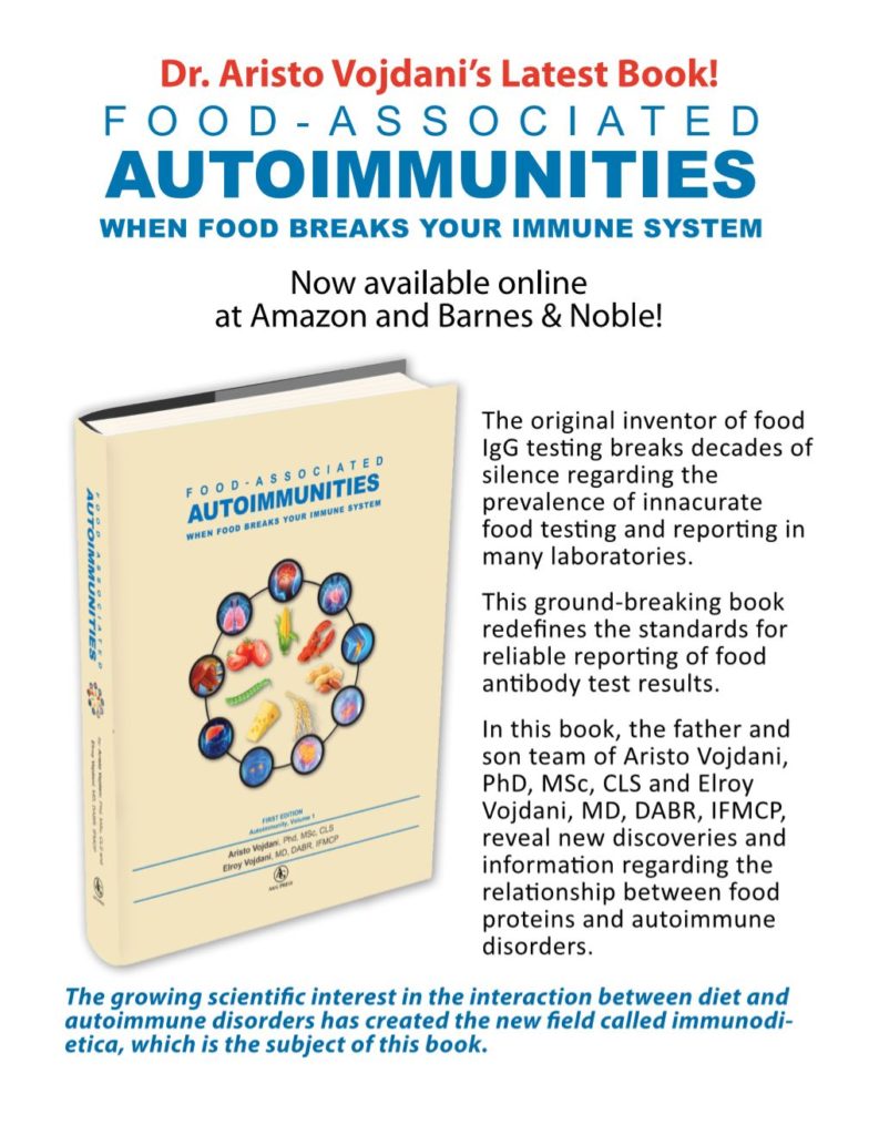
39. Vojdani A, editor. Food-associated autoimmunities: When food breaks your immune system. Los Angeles: A&G Press; 2019.
40. Gershteyn I, Ferreira L. Immunodietica: A data-driven approach to investigate interactions between diet and autoimmune disorders. J Translational Autoimmunity. April 2019.
41. Levin EC, et al. Brain-reactive autoantibodies are nearly ubiquitous in human sera and may be linked to pathology in the context of blood-brain barrier breakdown. Brain Res. 2010;1345:221–232.
42. Clifford PM, et al. Abeta peptides can enter the brain through a defective blood-brain barrier and bind selectively to neurons. Brain Research. 2007;1142(1):223-236.
43. Bredesen DE. Reversal of cognitive decline: a novel therapeutic program. Aging (Albany NY). 2014;6(9):707–717.
44. Bredesen DE, et al. Reversal of Cognitive Decline: 100 Patients. J Alzheimers Dis Parkinsonism. 2018;8:450.
45. Risk reduction of cognitive decline and dementia: WHO guidelines. Geneva: World Health Organization; 2019. Licence: CC BY-NC-SA 3.0 IGO.
Affiliation:
Immunosciences Lab., Inc.
822 S. Robertson Blvd., Ste. 312
Los Angeles, California 90035
Corresponding Author:
Aristo Vojdani
drari@msn.com
Tel: 310-657-1077
Fax: 310-657-1053


