Townsend Letter Alternative Medicine Magazine


  FREE e-Edition


 EDTA Chelation Therapy


 E-mail List


 TLDP.com


From the Townsend Letter
October 2018

A Mystery Answer to Restoring Brain Health
by David I. Minkoff, MD, Julie Mayer Hunt, DC, DICCP, FCCJP, and Ron Tindell
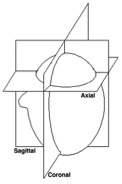
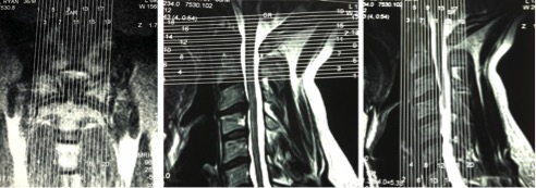
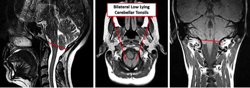
Search this site

Page 1, 2, 3

CCJ Imaging Methods
For CCJ MRI imaging, the benefits of upright versus supine imaging were discussed earlier in this article. For the recommended upright imaging, patients are seated; and images are obtained on the coronal, sagittal and axial planes (depicted in Figure 9) using sequences as shown in Figure 10.In these sequences:

  • The slice thickness in these cases should be a maximum of 2.8 mm.
  • The axial slices were obtained in proton density (PD), which is best to see ligaments.
  • The sagittal slices were obtained in T1 (longitudinal relaxation time) and T2 (transverse relaxation time).
  • Coronal images were obtained in T1.

BioDisruptUpright MRI imaging of the CCJ revealing low cerebellar tonsils in all three planes is shown in the Figure 11. This positioning may obstruct the normal flow of CSF. CFS obstruction may contribute to headaches, head pressure, dizziness, brain fog, and the like.

Figure 9

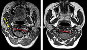
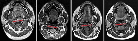
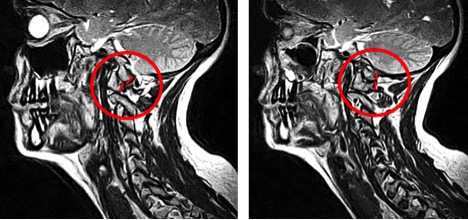
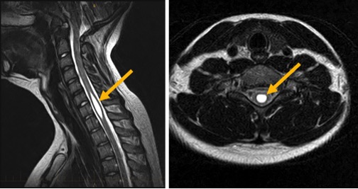
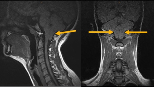
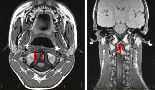
Figure 10

Figure 11

Atlas Rotation Observations: When the atlas rotates, it is plausible anatomically that the transverse process can abut the internal jugular vein. Figure 12 depicts two examples of atlas rotation misalignment. The red line highlights the rotation. The yellow arrow points to an internal jugular, which appears to have been compressed by the misaligned atlas. This compression can potentially affect venous outflow from the brain, causing backup of venous metabolic waste blood in the brain which is suggested in neurodegenerative brain diseases.10 Also note the football shape versus a normal, round shape of the spinal cord which plausibly can suggest dentate ligament attachment tension at the brainstem.5,11

Figure 12

C2 (Axis) rotation can be observed on CCJ oriented MRIs. Figure 13 provides several examples of axial rotational misalignment. The standard medical community cervical spine MRI misses this segment because the slices start at the C2/C3 disc. When one considers the vertebral artery pathway, illustrated in Figure 2, the axial misalignment can plausibly correlate with vertebral artery insufficiency and also the misalignments can affect dentate ligament tension of the spinal cord.5,11

Figure 13

C1 misalignment can be observed in the sagittal view with respect to the occipital condyles and the atlas lateral mass position. Figure 14a suggests anterior misalignment of the atlas lateral mass with respect to the occipital condyle. Figure 14b depicts a normal positioning of the C0/C1 articulation.13

Figure 14a and 14b

     
Observations that can be made through upright MRI have the potential to clearly objectify spinal misalignment (Subluxation) and clarify patient care needs. The CCJ is a vulnerable region and merits special consideration for care and treatment. There are many parameters for studying the CCJ through MRI which can range from CSF and blood flow impedance, ligamentous laxity and or insufficiency, and cerebellar tonsular ectopia as well as Chiari involvement.13
     
In 2012 the glymphatic system was postulated14 with regards to lymphatic drainage and brain health. The lymphatic system that was discovered in the brain is dependent on CSF flow. The glymphatic system, as shown in Figure 15, is a functional waste clearance pathway for the central nervous system. The CSF flow, when obstructed, appears to have negative plausible effects on brain health. Therefore, having the CCJ aligned contributes to non-obstructed flow of CSF and will plausibly contribute to improved brain health and immune function.

Figure 15

Craniocervical Syndrome Case Studies
Study 1, Pediatric Syrinx: A 14-year-old male presented in the office following ten days of hospitalization at a Johns Hopkins affiliated children's hospital facility for severe head, neck, and upper extremity pain and sensitivity. On release, it was explained to him and his family that hospital protocol had been exhausted and his pain was hormonal. MRI imaging inclusive of the CCJ was ordered the day after his hospital release, which revealed a large syrinx and cerebellar tonsillar ectopia (CTE) with CCJ misalignment. We postulate that the CCJ misalignment affected the CSF hydrodynamics and the misdirected CSF played a major role in formation of the syrinx. The MRI images in Figure 16 show the location and magnitude of the syrinx as well as showing the CTE.

Figure 16a and 16b

     
As treatment, CCJ realignment was performed and additional CSF flow imaging was obtained and utilized in the CCJ correction. The patient resolved successfully with this treatment.
     
Study 2, Chiari: A 19-year-old female presented to the office with eye-popping headaches, dizziness, and brain fog, which had been unremitting for the previous ten years since she had fallen on her head from a gymnastics uneven bar. She had an exhaustive list of neuro-medical consults which had provided no diagnosis or relief. She was told her issues "were all in her head." An MRI was ordered and revealed a Chiari of 22 millimeters as highlighted by the arrows in Figure 17.
     
She responded fairly well to CCJ specific care for three years, but ultimately had a decompression procedure that has been successful.

Figure 17

     
Study 3, Concussive Headaches: A 14-year-old male presented with constant severe head and neck pain following a concussion. MRI CCJ imaging was ordered, but the parents delayed obtaining the imaging for months. Ultimately, when the imaging was preformed, shown in Figure 18, a torn transverse ligament was discerned. The atlanto dental interspace is unequal (note the difference in spacing between the red marked and unmarked sides in the figure) therefore appropriate care inclusive of CCJ facet blocks are considered and possible CCJ stem cell therapy. CCJ instability is a strong consideration for his symptomatology, and the brainstem is football shaped due to dentate ligament traction parameters versus a normal round shape

Figure 18

Page 1, 2, 3


Consult your doctor before using any of the treatments found within this site.

Subscriptions are available for Townsend Letter, the Examiner of Alternative Medicine
magazine, which is published 10 times each year. Search our pre-2001 archives for further information. Older issues of the printed magazine are also indexed for your convenience.
1983-2001 indices ; recent indices. Once you find the magazines you'd like to order, please
use our convenient form, e-mail subscriptions@townsendletter.com, or call 360.385.6021.


360.385.6021
Fax: 360.385.0699
info@townsendletter.com

Who are we? | New articles | Featured topics | e-Edition |
Tables of contents
| Subscriptions | Contact us | Links | Classifieds | Advertise |
Alternative Medicine Conference Calendar | Search site | Archives |
EDTA Chelation Therapy | Home

© 1983-2018 Townsend Letter
All rights reserved.
Website by Sandy Hershelman Designs