Full article: Online publication only
Emil K. Schandl, MS PhD,FACB, SC (ASCP), CC (NRCC), LNC, CLD, is a clinical biochemist and oncobiologist with Metabolic Research, a 501(c)(3) not for profit biomedical research corporation, and American Metabolic Laboratories.
Abstract
The Cancer Profile (CA Profile) is a proposed adjunct diagnostic tool for early cancer detection and follow-up. The components are (1) tumor markers: HCG-Intact Hormone by IRMA, HCG-Total Hormone (intact, nicked intact, free β, nicked, and free β fragments) by chemiluminescence, HCG-Urine test by chemiluminescence, PHI enzyme (phosphohexose isomerase or glucose phosphate isomerase) by enzyme kinetics, and CEA (carcinoembryonic antigen) by chemiluminescence; and (2) peripherally related assays: GGTP, TSH, and DHEA-S. The CA Profile is neither organ nor site specific. It is designed to detect malignant neoplasms at their earliest stages, before other currently available diagnostic measures are efficacious. The CA Profile has proven to be an excellent adjunct tool for early detection of malignancies by producing abnormal clinical laboratory results, even years prior to actual diagnosis by current state-of-the-art methods. It is also of great value in monitoring the progress of cancer patients.
Introduction
Cancer is the number one killer disease of Americans when counting deaths related to pneumonia, heart failure, and so on caused by some treatment complications. In 2009, the Centers for Disease Control and Prevention attributed 559,888 deaths to cancers of all sites.1,2 Unfortunately, our healing arts specialists lack consistently effective weapons to combat the monstrous and many-faced disease.
Established traditional methods of varying chemotherapy regimens, radiation therapy, available immune therapies, and surgery lack the desired curative results. Optimally, the future of diagnostics and treatment is cure and not five-year survival. In addition, practitioners must strive to prioritize patient quality of life. The balance between quality of life and aggressive therapy is often tenuous; it behooves all practitioners to look to methods to stimulate the body's physiologic and immunologic participation, both in prevention and in treatment.
Many years may elapse during the progression of a normal cell to a cancerous one. Established traditional diagnostic methods are often too late to detect developing cancer early enough to substantially extend life. Palpation, X-ray, CT, MRI, PET, biopsy, and conventional tumor markers tend to reveal cancers already firmly established. At this point, the diagnosis is devastating to the patient; however, the cancer did not arise overnight. Environmental factors contribute to formation of many cancers. In fact, the patient may have been providing a tumor-coddling milieu – one that is alterable. Thus, the answer lies in prevention and ultra-early detection. In addition, following a patient's progress by repeating tumor markers may allow informed judgment of the success or failure of any patient-chosen therapy. The CA Profile has been instrumental in the management of patients with various cancers by a growing number of physicians of all disciplines, and thousands of patients with or without cancer. Herein, we will present several case studies and a brief review of the literature.
Historical Foundation
Dr. John Beard introduced a unifying theory of cancer origin as early as 1902.3 He hypothesized that cancers originate from embryonic cells, the trophoblasts. A major product of these cells is human chorionic gonadotropin hormone (HCG), the pregnancy hormone. Interestingly, HCG is present in a large percentage of all types of cancers of women, men, and children alike. In the 1950s, Dr. Manuel Navarro announced that he could detect HCG produced by minute tumor burdens – a few million trophoblast cells – using the H. H. Beard-Anthone urine test (BAT) for HCG.4 Since that time, research has shown that (1) HCG has both alpha and beta chains, and (2) HCG, FSH (follicle stimulating hormone), LH (luteinizing hormone), and TSH (thyroid stimulating hormone) have identical alpha subunits. Thus, the BAT is not specific for HCG. Improvements in technology have allowed investigators to better quantify specific HCG components for more accurate results.
In 1977, Dr. Robert R. Williams, in conjunction with the Framingham cholesterol study, reported the discovery of HCG hormone in the blood of study subjects two to three years prior to cancer diagnosis.5
The original 1980 Cancer Profile was designed with the premise that what one marker alone may miss, several together may not.6 The original Profile comprised three different tumor markers: HCG-β, PHI, and CEA. Utilizing the 1980 CA Profile, 94% of patients (n = 133) with proven cancers displayed at least one abnormal result. In addition, 26% of patients without proven cancer (n = 197) displayed one or more elevated tumor markers. A number of these "false positive" results were in individuals with signs or symptoms of cancer (e.g., enlarged lymph nodes, breast lumps, etc.), but without pathologic diagnoses. Some were later diagnosed with cancer.
From this investigator's perspective, "false positive" results, therefore, may represent an opportunity to positively and substantially influence the biophysical makeup of the patient – such as by promoting significant lifestyle changes. Such changes may lead to disappearance of the elevated marker and theoretically may result in cancer prevention. Alternatively, a "false negative" result may indicate a favorable treatment response. Thus, these markers should be followed regularly – prior to, during and following any patient-chosen therapy.
The CA Profile Markers
The current CA Profile consists of three tumor markers – HCG, PHI, and CEA with HCG measured by three complementary technical methods – and several markers of organ function (DHEA-S, TSH, GGTP). HCG is the pregnancy/malignancy embryonic hormone. PHI regulates cellular anaerobic metabolism and is the autocrine motility factor (AMF/malignancy factor). DHEA-S (dehydroepiandrosterone sulfate) is the adrenal antistress, proimmunity, and longevity hormone. TSH is an excellent screening assay for thyroid function. GGTP (gamma-glutamyl transpeptidase) monitors liver and other organ health.
- HCG: In 1987, Fujimoto et al. reported on an HCG-like substance (HCGLS) in the serum and tissue of patients with gastric and colorectal cancers.7 He assayed the serum and the tumors of his subjects simultaneously, and he concluded that the defective HCG; that is, aberrant HCGLS, is a characteristic of malignant tumors. Later researchers described the infrequent, low-level presence of pituitary HCGLS substance in older female patients.13 Pituitary HCG has an N-linked sugar side chain with more resemblance to luteinizing hormone (LH) than HCG. The researchers postulated that GRH (gonadotropin releasing hormone) stimulates the production of LH, FSH, and the HCGLS molecule. High doses of oral progesterone treatment for 3 weeks suppressed pituitary production of HCG. Thus, a progesterone "challenge" with resultant disappearance of HCG ruled out the presence of tumor-generated HCG.8 Pituitary HCGLS has significant biological activity12; thus, bioidentical hormone replacement therapy may be an answer for asymptomatic, LH and FSH elevated individuals in order to suppress pituitary HCGLS hormone production.9-11
The HCG hormone test is an accepted tumor marker for germinal cell tumors. However, volumes of biomedical literature have convincingly established that it is an excellent cancer indicator for most if not all types of cancers. Using 85 different cancer cell lines, Acavedo et al. described the expression of complete HCG in cell membranes in association with metastatic aggressiveness of tumors of different histological types and origin. Intact HCG was undetectable in benign cells. The researchers maintained that synthesis and expression of HCG, its subunits, and its fragments is a common biochemical denominator of cancer. They found translatable HCG-β mRNA in all the tested fetal and cancer cell lines. In fact, the authors reported detectable levels of HCG in the blood and urine of patients with cancers of the breast, bladder, gastrointestinal tract, lung, and skin (melanoma) as well as in embryonal carcinomas.16,17 In another study, the presence of the hormone was highly indicative of malignant, aggressive tumors of multiple sites including lung, pancreas, and liver.18-20 Clinical laboratory evidence obtained by performing ultra sensitive HCG tests at American Metabolic Laboratories (AML) also indicates that HCG might be present in all types of cancers.
In addition to acting as a marker for possible disease, HCG and HCGLS exhibit immune inhibition, angiogenesis promotion, and stimulation of metabolic, regulatory, growth and cell proliferation functions in target cells and organs. Thus, the presence of any quantity of HCG can potentially herald and contribute to the development and progression of any type of cancer.
Although some researchers (e.g., ClinLabNavigator.com) are concerned with the validity and clinical utility of low-level HCG in the absence of secondary evidence of disease or pregnancy, this investigator is more interested in advancing an epoch of previsual disease markers. Indeed, case studies indicate that the CA Profile does just this.
To establish the biological reality and identification of positive HCG results, multiple complementary assays are necessary. The CA Profile utilizes the following: (1) two positive technologically different serum HCG quantitative tests (e.g., HCG-IRMA, calibrated to ≥ 0.3 mIU/mL and HCG-IMM, chemiluminescence calibrated to ≥0.2 mIU/mL) and (2) urine confirmatory quantitative test by chemiluminescence, calibrated to ≥0.2 mIU/mL
- PHI: Phosphohexose isomerase, EC 5.3.1.9, is also known as glucose phosphate isomerase or phosphoglucose isomerase, and was originally isolated from a human melanoma cell line. PHI is a small GTPase of the Rho family with effects on cell growth, morphogenesis, cell motility, cytokinesis, trafficking and organization of cell cytoskeleton, cell motility, transformation, and metastasis. It also promotes angiogenesis by direct effects on endothelial cells. It is somehow involved in the accumulation of ascites and displays an antiapoptotic effect.14 It is a regulatory catalyst of anaerobic sugar metabolism (Embden-Meyerhof glycolytic and glucogenetic pathways) by reversibly converting glucose-6-phosphate to fructose-6-phosphate. Various authors designate this neurokine as the autocrine motility factor or AMF. It stimulates cell motility in an autocrine manner and is closely related to malignancy. PHI may be elevated in patients with malignant gastrointestinal, kidney, breast, colorectal, and lung tumors. This investigator postulates that a specific enzyme inhibitor may be developed or discovered that would prevent the negative effects of this enzyme.
Studies suggest that cancer cells may metastasize earlier than previously thought, although the mechanism is not yet apparent.15 This investigator hypothesizes that the biochemical initiator of metastasis is PHI, autocrine motility factor. Elevated plasma levels may lead to metastatic events: cytokinetic vibration and dislodgement of the cancerous cell from its neighboring environment and consequent embolism via lymph or blood flow to distant sites. American Metabolic Laboratories began manufacture of the PHI assay reagents when the previous manufacturer ceased production.
The PHI enzyme and HCG hormone tests have been successfully implemented and perfected as tumor markers. However, the tests are not FDA approved for such use. Doctors may use and apply the technology with watchful eyes as an experimental endeavor that may bring useful information to the forefront.
- CEA is a broad-spectrum tumor marker that may detect or monitor most malignant processes. It is an excellent marker for breast cancer.
- GGTP is a constituent of the profile for monitoring primarily hepatic, and renal, cardiac organ/tissue health.
- TSH directly affects thyroid function and thus, governs the body's basic metabolic rate; that is, rate of oxidative phosphorylation. A hypothyroid condition may usher in anaerobic glucose fermentation, postulated to lead to cancer. In addition, cancer therapies may evoke hypothyroidism.21,22
- DHEA is considered the adrenal antistress, proimmunity, and longevity hormone by this author. Studies conducted at AML indicate that most, if not all, cancer patients present with low DHEA levels. Often a 40-year-old individual may present with DHEA quantities of an 80-to-90 year old patient. The importance of this steroid hormone in cancer prevention and therapy should be noted.23,24
- Additional patient-indicated tumor marker studies may include the following: CA 19-9, CA 15.3, CA 125, and PSA (prostate specific antigen).
Assay Interpretation
Applicable Normal Values |
HCG-IRMA |
<1.0 |
mIU/mL |
HCG-IMM |
<1.0 |
mIU/mL |
HCG-Urine |
1.0–3.8 |
mIU/mL |
PHI enzyme |
0–34 |
U/L |
CEA |
0–3.0 |
ng/mL |
CA 19-9 |
0–37 |
U/mL |
CA 15.3 |
7.5 - 53.0 |
U/mL |
CA 125 |
1.9–16.3 |
U/mL |
PSA 3rd Gen |
0.00–2.80 |
ng/mL |
Figure 1 is an aid for the interpretation of the triple-HCG test assays utilizing the Three-Test criterion. Notably, none of the three methods detects the a-subunit molecular species shared among HCG, TSH, and LH. Instead, the HCG-specific β-subunit is measured.
Figure 1: Interpretation of HCG Results Utilizing the Three-Test Criterion
HCG-IRMA |
HCG-IMM |
HCG-Urine |
Interpretation |
− |
− |
− |
Negative |
− |
+ |
− |
Negative/Uncertain*** |
− |
− |
+ |
Negative/Uncertain*** |
+ |
− |
− |
Negative/Uncertain**** |
+ |
+ |
− |
Positive/Suggestive**** |
+ |
− |
+ |
Positive |
+ |
+ |
+ |
Positive |
− |
+ |
+ |
Positive/Suggestive ** |
**HCG-IMM/Urine may contain HCGLS;
*** Needs confirmatory urine or another Pos method;
****To confirm requires Pos Urine, especially if the patient has a protracted exposure to domestic animals. The IRMA test does not detect HCGLS, nor beta or fragments of tumor-generated HCG.
Uncertain/Suggestive results require clinical correlation.
Case Studies
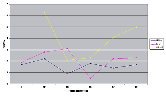
A 61-year-old female, "Susan," presented with a long history of gastric discomfort and pain of unknown etiology. The CA Profile (Figure 2) showed a consistent presence of HCG over the observed 19 months. Concern for a developing or already existing malignancy was prompted.
Figure 2: Susan 61 F, Gastritis, No Cancer Diagnosis
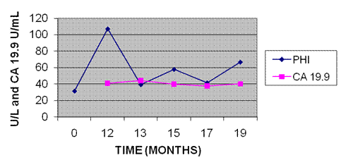
The PHI enzyme remained markedly elevated throughout the study period as well (Figure 3). Additional patient-specific studies were undertaken and the gastric/pancreatic tumor marker, CA 19.9, was marginally elevated, suggestive of gastritis. At this point in her medical workup, endoscopy, colonoscopy, and GI exams were negative. Cautious outlook for the patient's future and introduction of substantial lifestyle changes for cancer prevention were undertaken.
Figure 3: Susan 61 F, Gastritis
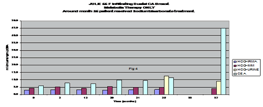
A 55-year-old female, "Julie," was diagnosed with infiltrating ductal breast carcinoma; no metastatic lesions were identifiable. A CA Profile was requested and the following results seen: HCG measurements remained elevated throughout the 37-month study period, but PHI remained within normal range (Figure 4). During the 35th month of the study, she traveled abroad for sodium bicarbonate tumor injections. She reported for follow-up evaluation at the 37th month; a dramatic increase of CEA was observed. At that point, she initiated herbal therapy, followed by a more successful course of chemotherapy (Herceptin, Tykerb, Femarra, and Zometa) at the Burzynszky Clinic in Houston, Texas.
A follow-up CA Profile is timely at this time.
Figure 4: Julie, 55 F
A 65-year-old male, "Charlie," presented for prostate discomfort. Two series of CA Profiles and Third Generation PSA tests were performed. At the beginning of the study, the CA Profile tests, with the exception of low DHEA-S, were within normal range (Figure 5). The picture changed 10 months later. The three HCG tests became marginally elevated, and the PHI test became substantially elevated. Charlie refused biopsy in fear of a malignant diagnosis and a possibly aggressive therapeutic procedure, and instead chose holistic metabolic therapy. Similarly to Charlie's case, with the aid of the CA Profile, a physician may be able to infer the presence of disease even in the absence of invasive procedures; patient-chosen treatments may be monitored for efficacy.
Figure 5: CA Profile of Charlie, 65-Year-Old Male with Prostate Discomfort
Date |
HCG-IRMA |
HCG-IMM |
HCG-Urine |
PHI |
PSA |
CEA |
TSH |
GGTP |
DHEA-S |
8/17/2008 |
0.1 |
0.1 |
1.2 |
28.7 |
2.09 |
0.9 |
1.26 |
10 |
155.0 |
5/30/2009 |
1.3 |
1.3 |
1.7 |
62.8 |
1.56 |
0.4 |
0.91 |
15 |
172.0 |
|
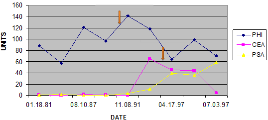
A 56-year old male, "Karl," reported for clinical laboratory evaluation for preventative reasons and without symptoms or complaints. He requested the CA Profile and Third Generation PSA tests (Figure 6). Initial results were negative for all the tests, with the exception of the PHI enzyme, which was markedly elevated. He was reexamined periodically. The enzyme remained elevated for 16 years, the entire length of the study. At 10 years and 10 months, Karl underwent prostate biopsy. Metastatic prostate cancer was diagnosed (first arrow, Figure 6). At this point, the CEA and PSA tumor markers also began to rise. Six years later, in the light of continuous CEA elevations, a second primary cancer site, colon cancer was identified and resection was undertaken (second arrow, Figure 6). The CEA returned to normal following surgery; however, the PHI and PSA remained elevated. After intensive chemotherapy and radiation, all in vain, Karl returned his spirit to his Creator.
Figure 6: Karl, 56 M CA Prostrate 10-Year History
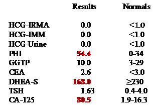
A 46-year-old female, "Sheila," presented with an initial diagnosis of ovarian cysts and uterine fibroids. She requested a CA Profile and CA-125, the traditional ovarian tumor marker. Markedly elevated PHI and CA-125 suggested the development or presence of metastatic ovarian disease (Figure 7). DHEA, as noted earlier, was depressed as in most cancer patients and those developing cancer of any sort (investigator observations).
Figure 7: Sheila F 46, Ovarian Tumor, Uterine Fibroids
A 72-year-old female, "Earleen," presented with a history of infiltrating ductal breast carcinoma and 4+ breast thermogram. HCG was elevated by two separate assays. PHI and CEA were also elevated (Figure 8). Elevated PHI in this case was concerning for possible metastasis. The low DHEA-S level may have indicated insufficient immune status. Therefore, the presence of malignant breast cancer may have generated the high-grade positive thermogram. As noted earlier, the CA Profile is not site specific; however, a thermogram could map out the whereabouts of a tumor.
Figure 8: Positive Thermogram Confirmed by the CA Profile
HCG-IRMA |
0.0 |
HCG IMM |
1.7 |
HCG Quant Urine |
2.3 |
PHI |
56.6 ↑ |
GGTP |
15.00 |
CEA |
12.2 ↑ |
DHEA-S |
52.4 ¯ |
TSH |
1.47 |
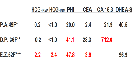
Figure 9 depicts three outcomes of three different patients who chose to utilize the CA Profile: (1) no cancer, negative results; (2) no cancer diagnosis, positive results; and (3) cancer diagnosis, positive results. P. A., a 49-year-old female, presented with no cancer diagnoses and normal CA Profile. D. P., a 36-year-old female, also had no diagnosed abnormalities, yet presented with elevated PHI and grossly elevated CA 15.3. E. Z., a 52-year-old female, presented with a diagnosis of infiltrating ductal carcinoma of the breast and the four tumor markers tested were elevated. Patient D. P.'s clinical laboratory results clearly indicated a developing or already existing breast cancer. The site specificity determination became obvious from the rather high, traditional CA 15.3 test. Of note, the CA 15.3 test is only a viable tumor marker when it is positive. This may be ~2% of the time, whereas the CA Profile positive predictive value can be as high as 93% to 98%.25
Figure 9: Utilization of the CA Profile
CA Br Case History Examples
Patient Series
- Breast Cancer: 92% of 240 patients with pathologically established breast cancer patients yielded elevated CA Profile markers (Figure 10).
- Colon cancer patients: 93% (n = 59) yielded elevated CA Profile values (Figure 11).
- Lung cancer patients: 92% (n = 129) showed 1 to 3 positive tumor markers (Figure 12).
Figure 10: Breast Cancer Study
Elevated Markers |
n |
% |
|
(HCG, PHI, CEA) |
|
|
|
0 |
19 |
8 |
|
1 |
54 |
22 |
92% |
2 |
107 |
45 |
Positive |
3 |
60 |
25 |
Markers |
Total |
240 |
100 |
|
Figure 11: Colon Cancer Study
No. Elevated Markers |
n |
% |
|
HCG, PHI, CEA |
|
|
|
0 |
4 |
7 |
|
1 |
13 |
22 |
93% |
2 |
30 |
51 |
Positive |
3 |
12 |
20 |
Markers |
Total |
59 |
100 |
|
Figure 12: Lung Cancer Study
| No. Elevated Markers |
n |
% |
|
HCG, PHI, CEA |
|
|
|
0 |
2 |
3 |
|
1 |
23 |
18 |
98% |
2 |
46 |
35 |
Positive |
3 |
58 |
44 |
Markers |
Total |
129 |
100 |
|
Discussion
The Cancer Profile has proven to be an excellent adjunct clinical laboratory medicinal method for early detection as well as follow-up of cancers of all types. Because of the viability and powerful clinical utilization, an ever-growing number of physicians and indeed patients are using it to their satisfaction. The documentations here presented are only a small fraction of available literature on the matter of the individual tests. The CA Profile has passed the test of time, remaining a viable diagnostic tool for almost three decades with several interval technical improvements. In the 1970s, this investigator developed anti-HCG antibodies in rabbits at the Howard Hughes Research Institute in Miami. Since then, ever more sensitive, accurate, and precise tests became available and used with some in-laboratory modifications to create the current CA Profile. The PHI enzyme test was available commercially for several years. The manufacturer discontinued it for lack of adequate commercial interest. Dr. Schandl requested and received the formula, and now with a few modifications is producing it with the support of Metabolic Research, a 501(c)(3) not-for-profit corporation. It should be noted that detection of PHI, even without evidence of a tumor, may identify a new subset of micrometastasis, to identify malignancy even more focal than the current definition (i.e., cell clusters larger than 0.2 mm, or isolated tumor cells [circulating tumor cells?] smaller than 0.2 mm cell clusters. See 7th edition of the AJCC Cancer Staging Manual and CAP Today, March 2010). The question may be posed, does PHI, the autocrine motility factor and neurokine, initiate micrometastasis before wandering malignant cells can be detected even by a microscope? Obviously, the triad of HCG hormones and the PHI enzyme are the backbones of the CA Profile. However, for the most robust clinical laboratory evaluation, the entire profile is optimal.

Notes
- Centers for Disease Control. Leading causes of death [Web page].
www.cdc.gov/nchs/fastats/lcod.htm.
- American Cancer Society. Cancer facts and figures. 2009.
- Beard J. The Unitarian trophoblastic theory of cancer. Lancet. 1902;1:1758.
- Navarro M. 72nd Sci Meeting of the Cavite Med Soc; 1959; Trece Martires City, Philippines.
- Williams RR, McIntire KR, Waldmann TA, et al. Tumor-associated antigen levels (CEA, HCG, alpha-feto protein) antedating the diagnosis of cancer in the Framingham study. J Natl Cancer Inst. 1977 June;58(6):1547–1551.
- Schandl EK. Clinical biochemical parameters in cancer diagnosis and therapy. Clin Chem. 1980;26(7):1040.
- Fujimoto S et al. The presence of an aberrant type of human chorionic gonadotropin in patients with gastric or colorectal cancer. Jpn J Surg. 1987 Sep;17(5):382–387.
- Cole LA. Background human chorionic gonadotropin in healthy, non-pregnant women. Clin Chem. 2005;51(10):1765–1766.
- Birken S et al. Isolation and characterization of human pituitary chorionic gonadotropin. Endocrinol. 1996;137:1402–1411.
- Stenman UH et al. Serum levels of HCG in non-pregnant women and men are modulated by GRH and sex steroids. J Clin Endocrinol Metab. 1987;64:730–736.
- Gronowski A et al. Use of serum FSH to identify perimenopausal women with pituitary HCG. Clin Chem. 2008;54(4):654–656.
- Birken, S. et al. Isolation and Characterization of Human Pituitary Chorionic Gonadotropin. Endocrinology 1996; 137: 1402-1411.
- Cole LA, Khanlian SA. Inappropriate management of women with persistent low HCG results. J Reprod Med. 2004;49:423–432.
- Ynagava T et al. Novel roles of the autocrine motility factor/phosphoglucose isomerase(PGI or PHI) in tumor malignancy. Endocr Relat Cancer. 2004;11(4):749–759.
- Daily Diagnosis, Leading News, American Society for Clinical Pathology. August 29, 2008.
- Acavedo HF et al. Flow cytometry method for the analysis of membrane-associated HCG, its subunits and fragments on human cancer cells. Cancer. 1992; 69:1818–1828.
- Acavedo HF et al. HCG-β gene expression in cultured human fetal and cancer cells. Cancer. 1995;76:1467–1475.
- Marcillac I et al. Free human chorionic gonadotropin β subunit in gonadal and nongonadal neoplasms. Cancer Res. 1992;52:3901–3907.
- Martinez Flores A et al. Development and validation of an in vitro culture model for the study of the differentiation of human trophoblast. [In Spanish.] Ginecol Obstet Mex. 2006 Dec;74(12):657–665.
- Bjurlin MA et al. Histological pure seminoma with an elevated beta-HCG of 4497IU/l. Urology. 2007 Nov;70(5):1007.e13–1007.e15.
- Smith G L, et al. Risk of hypothyroidism in older breast cancer patients treated with radiation. Cancer. 2008 Mar 15;112(6):1371–1379.
- Madanat LM et al. Hypothyroidism among pediatric cancer patients. Int J Cancer. 2008 Apr 15;122(8):1868–1872.
- Oberbeck R et al. Dehydroepiandrosterone: a modulator of cellular immunity and heat shock protein 70 production during polymicrobial sepsis. Intensive Care Med. 2007 Dec; 33(12):2207–2213. Epub Sep 2007.
- Rook GA, Baker R, Zumla A. Steroid metabolism and immunity: therapeutic implications. BioDrugs. 1997 Sep;8(3):157–163.
- Schandl EK, Carlo A, Schandl CA. Cancer profile v. CA 15.3 tumor marker [abstract]. Am Assoc Bioanalysts, 50th Anniversary Conference; Las Vegas, NV; 2006 Jun.
|


![]()
![]()
![]()


